Tag Archives: cell

Gene variant may provide novel therapy for several cancer types

This landmark study is published in the June 6, 2013 issue of the journal Science. Ramon Parsons, MD, PhD, Professor and Chair of Oncological Sciences led the team that discovered a mutation in the tumor suppressor gene PTEN, which has subsequently been recognized as the second most common mutation in cancer, especially in breast, prostate, and brain cancers. PTEN encodes a 403 amino acid lipid phosphatase protein that is critical to cellular growth, proliferation, and survival…

More research needed on anesthesia’s impact on brain, study shows

Surgical anesthesia’s impact on the brain has long been debated, and even anesthesiologists have admitted the effects of these drugs on humans is not clearly understood. “Anesthetics have been somewhat of an enigma; nobody knows how they really work, and we basically use them in thousands of patients every day,” study author Dr. Andreas Loepke, a physician and researcher in the department of anesthesiology at the Cincinnati Children’s Hospital Medical Center, told FoxNews.com. Concerns have been raised among anesthesiologists like Loepke over previous research indicating that exposure to anesthesia may increase the rate of cell death in the brains of young animals. And now, a new study in mice published in Annals of Neurology indicates that anesthesia seems to kill off younger neurons more often than older neurons – regardless of the age of the animal.  Researchers have not yet studied the impact of anesthesia on human brain cells. “You can’t section a human brain,” Loepke said. “…But if it were occurring in humans, we would predict that anesthetics affect neurons in patients of all ages.” Loepke and his colleagues examined the rates of cell death in the brains of mice exposed to anesthesia for six hours. They focused particularly on the dentate gyrus region of the brain, which helps control learning and memory. “We found something very interesting, in that cell death occurred in the spot where the dentate gyrus forms new neurons,” Loepke said. The root cause and impact of anesthesia-related cell death is unknown, and the study’s authors said more research needs to be done.   “During development, (we) form twice as many neurons as we need as an adult. The brain needs to be pruned back to properly function,” Loepke said. “So it’s currently unknown whether anesthesia kills neurons that would have been eliminated anyways from the brain or neurons later needed for vital function.”   Loepke added that human studies of older adults have indicated that some people do experience memory problems after undergoing anesthesia, which can be short-term or long-term.  Whether this is caused by anesthesia or by the body’s reaction to pain or surgery remains unclear. “The need for surgery could be a marker for these problems occurring – (or the) inflammatory response to the body from the surgery. These have all been found to alter neurons,” Loepke said. Loepke and his colleagues hope to go on to study the effect of anesthesia in humans using magnetic resonance imaging (MRI) scans. But until more research is done, Loepke urged people not to worry too much. “Patients need to make sure they get the surgery they need, because putting off the surgery could put you at more risk (than the anesthesia),” Loepke said.source : http://www.foxnews.com/health/2013/06/05/more-research-needed-on-anesthesias-impact-on-brain-study-shows/

Errors in cloning study cast doubt on publication process

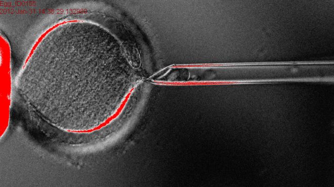
A headline-making paper last week announcing that scientists had, for the first time, cloned human embryos and harvested stem cells from them contains minor errors, the authors acknowledged on Thursday.  The mistakes raised questions about how well the journal that published the paper vetted it but probably do not undermine the study's central claim. In a statement, the journal, Cell, said “there were some minor errors” in the paper, but “we do not believe these errors impact the scientific findings of the paper.” An anonymous commenter on the website PubPeer, where scientists discuss papers after they have been published, first pointed out problems with the paper, which drew extensive media coverage. Even before the errors were spotted, however, there was concern among experts not involved in the study that Cell had rushed publication. It received the manuscript on April 30, tapped outside scientists to review it in the standard process called peer review, asked the authors to make revisions based on that review and accepted the paper on May 3. When asked about the short turnaround time last week, Cell spokeswoman Mary Beth O'Leary said the paper “underwent a rigorous peer review and editorial process.” Outside experts disagree. A three-day review process “is almost impossibly fast,” said cell biologist Jim Woodgett of Mount Sinai Hospital in Toronto, Canada. “To have a paper like this received, reviewed, revised and accepted so quickly is very, very unusual.” In a statement on Thursday, Cell referred to “the preeminence of the reviewers” (whom it would not identify) and said it has “no reason to doubt the thoroughness or rigor of the review process.” The rapid turnaround was possible because the reviewers “graciously agreed to prioritize” the paper. DOLLY REDUX The paper described how scientists led by biologist Shoukhrat Mitalipov of Oregon Health & Science University accomplished what others had failed to: “therapeutic cloning” in humans. That procedure begins with a human egg. The Oregon scientists removed its genetic material, or DNA, then took an adult skin cell and fused it with the egg. The DNA in the skin cell took over, causing the egg to begin developing as if it had been fertilized. This “somatic cell nuclear transfer” was used to clone Dolly the sheep in 1996. But in this case, the goal was not a human being; Mitalipov said last week that scientists would not implant the dividing embryo into a womb so that it could develop into a baby. Instead, the aim was a dishful of stem cells, which can morph into any of the 200-plus cells in the human body and might be used therapeutically, such as to replace cells lost to degenerative diseases such as Parkinson's. After a few days, the human embryo contained exactly that: stem cells that the Oregon scientists could use to start cell lines. The errors in the Cell paper involve photographs and data plots, something OHSU spokesman Jim Newman said was “an editing error, not issues with the research or the data itself. “OHSU agrees that there were some minor errors made when preparing the figures for initial submission,” Newman said, adding that the university does not believe the errors “impact the scientific findings of the paper in any way. We also do not believe there was any wrongdoing.” In one case, an image described as a cloned stem-cell colony is reproduced in another image, where it is labeled an embryonic stem-cell line derived from in vitro fertilization (IVF), not cloning. Mitalipov told the journal Nature that the label is wrong, and that another labeling mistake explained other duplicated images. Another error was in images purporting to show that the genes that are turned on in stem cells derived from the cloned embryo (such as genes that make a cell a neuron) are similar to those in stem cells taken from IVF embryos, considered the gold standard for embryonic stem cells. The point was that the stem cells taken from the cloned embryos are true stem cells. The problem, said the anonymous reviewer on PubPeer, is that the two images - genes activated in IVF stem cells and in clone stem cells - are suspiciously identical. Mitalipov said one image used the wrong data, and that he and his team are correcting it. While the mistakes seem innocent, they raised concerns among stem-cell researchers because the field has been struck by fraud in the past. In 2004 scientists led by Woo Suk Hwang of Seoul National University claimed to have produced human embryonic stem cells through the same technique used by the Oregon team. Their paper, published in Science, turned out to contain fabricated data. That came to light when scientists figured out that some of the images in the paper were copied or manipulated. “When I read the Hwang paper, I didn't find any glaring problems” at first, stem-cell biologist George Daley of the Harvard Stem Cell Institute said, explaining how difficult it is to spot fraud. “I am waiting to learn more, but there is a difference between errors in photomicrographs and fraudulent production of cell lines,” he said. So far, most scientists' ire is being directed at Cell more than the Oregon researchers. “To thoroughly evaluate the claims requires delving into the data, and you can't expect people to do that in a day or two,” said Mount Sinai's Woodgett, referring to peer review. “You're forcing them to be superficial.” Science journals compete intensely for “hot” papers, which can translate into headlines, subscriptions and advertising. Cell is published by Elsevier, a division of Reed Elsevier . Six years ago, Nature held up by six months a paper by Mitalipov in which his team used the Dolly method to clone monkey embryos, the journal reported on Wednesday. Scientists sometimes shop around hot papers, seeking a journal that will publish it fastest. Mitalipov told Nature he was in a hurry to get his Cell paper out before a stem cell meeting in June.source : http://www.foxnews.com/health/2013/05/24/errors-in-cloning-study-cast-doubt-on-publication-process/
Research identifies a way to make cancer cells more responsive to chemotherapy

Dr. Shawn Li, PhD, and his team at Western’s Schulich School of Medicine & Dentistry, identified that a protein called Numb functions to promote the death of cancer cells by binding to and stabilizing a tumor suppressor protein called p53 -a master regulator of cell death. The scientists found when Numb is reduced or methylated by an enzyme called Set8, it will no longer protect p53. …

When oxygen is short, EGFR prevents maturation of cancer-fighting miRNAs

Under conditions of oxygen starvation often encountered by tumors, the epidermal growth factor receptor (EGFR) gums up the cell’s miRNA-processing machinery, an international team led by scientists at The University of Texas MD Anderson Cancer Center discovered. "So when hypoxia stresses a cell, signaling by EGFR prevents immature miRNAs from growing up to fight cancer," said senior author Mien-Chie Hung, Ph.D., professor and chair of MD Anderson’s Department of Molecular and Cellular Oncology and holder of the Ruth Legett Jones Distinguished Chair…

Cold plasma successful against brain cancer cells, study suggests

If someone is diagnosed with the type of brain tumour called glioblastoma, the prospects are dire: median survival is just a bit over one year, and less than 16 % of the patients survive more than three years. It is still unknown how this cancer is triggered — only a few rare genetic factors have been identified so far — and treatment remains largely palliative, i.e. trying to alleviate the symptoms and prolonging the life of the patient. …

How gold nanoparticles can help fight ovarian cancer

The discovery is detailed in the current online issue of the Journal of Biological Chemistry. "This study identifies a novel mechanism that protects ovarian cancer cells by preventing the cell death or apoptosis which should occur when they encounter positively charged nanoparticles," say the senior authors of this study, Priyabrata Mukherjee, Ph.D., a Mayo Clinic molecular biologist, and Y. S. Prakash, M.D., Ph.D., a Mayo Clinic anesthesiologist and physiologist…

In medical breakthrough, scientists convert human skin cells into embryonic stem cells

In a major medical breakthrough, researchers at the Oregon National Primate Research Center (ONPRC) have for the first time ever successfully converted human skin cells into embryonic stem cells – via a technique called nuclear transfer. The research has major implications for the future of medical treatments, as many believe embryonic stem cells are the key to treating damaged cells lost through injury or illness.  According to various medical researchers, stem cell therapy has the potential to treat anything from heart disease and spinal cord injuries to major neurological diseases, like Parkinson’s disease and multiple sclerosis. Through a common laboratory method known as somatic cell nuclear transfer (SCNT), ONPRC scientists, along with researchers at Oregon Health & Science University, essentially swapped the genetic codes of an unfertilized egg and a human skin cell to create their new embryonic stem cells.  The researchers were able to fine-tune this method through a series of studies in macaque monkeys, and this study marks the first successful use of the SCNT technique to create embryonic stem cells in humans after many failed attempts from other research laboratories around the world. The SCNT process involved scooping out the nucleus of a donated egg cell, leaving nothing by the egg’s cytoplasm – an essential ingredient.  The researchers replaced the egg’s nucleus with the nucleus of a skin cell, which contains an individual’s genetic code.  The combination of the egg’s cytoplasm and the skin cell’s nucleus eventually grows and develops into the embryonic stem cell. “The idea is that the egg cytoplasm has some factors – we don’t know their nature yet – but it has the ability to reset the cell’s identity,” lead author Dr. Shoukhrat Mitalipov, a senior scientist at ONPRC, told FoxNews.com.  “It basically erases all this memory, and now we can derive them and make them into stem cells.” Embryonic stem cell research has been a controversial pursuit since its conception.  Derived from unused embryos, which have been fertilized in vitro, these types of cells are pluripotent – meaning they have the capacity to develop into a variety of different cell types within the human body. However, the utilization of embryonic stem cells has been met with ethical concerns, as many believe a fertilized egg should be granted the status of a human being.  Because the fertilized human embryo is ultimately destroyed in the development of embryonic stem cells, many activist groups and politicians have spoken out against the use of such medical techniques. Fortunately, the SCNT method bypasses these ethical dilemmas, as the donated eggs are never actually fertilized.  Instead, the researchers ‘trick’ the egg cell into thinking it has been fertilized by a sperm.  During the nuclear transfer process, Mitalipov and his team prompt the cell to remain in a state known as “metaphase” – a stage of cell division in which the cell’s chromosomes align in the middle of the cell just before the cell divides.  This keeps the process from stalling and encourages the cell to ultimately develop into a stem cell. Once reprogrammed, these stem cells can be cultivated into any kind of living cell – including nerve cells, heart cells, brain cells and many more. Mitalipov also noted that once an individual has this process done, he or she will have a lifelong “bank” of embryonic stem cells doctors can utilize whenever the patient is injured or sick. “You only need to do it once per patient,” Mitalipov said.  “Once established, the stem cells are like a permanent source of stem cells – they have an unlimited capacity to keep growing…  We only need to do it once, but one colony multiples to make hundreds of colonies.  We can freeze them and continue to grow more and more.” The researchers addressed the possibility that the SCNT method may be considered therapeutic cloning, but they said it is highly unlikely this method would be able to produce human clones.  In all the years of utilizing SCNT with monkey cells, no monkey clones have ever successfully produced, so it is highly unlikely that human clones can be produced through this method – especially since human cells are much more fragile than monkey cells. Instead, Mitalipov hopes people focus on the main goal of the research, which is to foster hundreds of different studies utilizing this new process of developing embryonic stem cells. “We still have a lot more work to do, to learn how to transplant them,” Mitalipov said. “But the first step is already done, and it looks pretty clean.  Probably, people could start banking these cells now; and hopefully, in the near future we will have some treatments coming in the clinic.” The research was published online in the journal Cell.source : http://www.foxnews.com/health/2013/05/15/in-medical-breakthrough-scientists-convert-human-skin-cells-into-embryonic-stem/